
Cutaneous lesions across the face, head, and neck present both diagnostic and aesthetic considerations. While the majority of facial moles (nevi), epidermoid cysts, and seborrheic keratoses are benign, sun-damaged skin across South Florida exhibits elevated rates of pre-cancerous actinic keratoses, basal cell carcinomas, squamous cell carcinomas, and melanoma. At Oral and Facial Surgery of Miami in Coral Gables, board-certified oral and maxillofacial surgeon Dr. Johanny Caceres performs surgical excisions and biopsies designed to guarantee clear margins while minimizing visible facial scars.
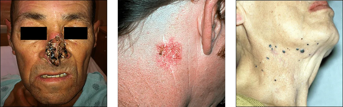
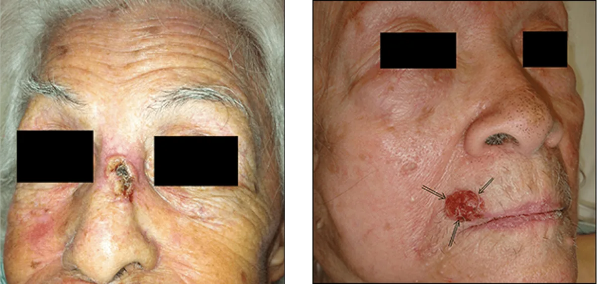
Cutaneous Pathology and Clinical ABCDE Diagnostic Criteria
Differentiating harmless pigmented blemishes from aggressive malignancies requires systematic clinical inspection using dermoscopy and established ABCDE diagnostic guidelines: Asymmetry (one half unlike the other), Border irregularity (scalloped, notched, or poorly defined margins), Color variation (shades of brown, black, red, or white), Diameter (exceeding six millimeters), and Evolving characteristics (changes in size, shape, bleeding, or itching).
Basal cell carcinoma (BCC) represents the most common non-melanoma skin cancer, presenting classically as a pearly, translucent papule with prominent arborizing telangiectasias (spider veins) and rolled edges. Squamous cell carcinoma (SCC) presents more aggressively as an indurated, scaling plaque or non-healing ulcer with metastatic potential. Melanoma requires immediate, decisive intervention because depth of invasion (Breslow thickness) correlates directly with systemic prognosis.
Biopsy Modalities: Shave, Punch, Incisional and Excisional Techniques
When a cutaneous lesion presents suspicious clinical characteristics, an appropriate biopsy must establish histological identity before large-scale surgery. A tangential shave biopsy removes a superficial epidermal disc, useful for elevated benign keratoses or suspected superficial basal cell lesions. A circular punch biopsy obtains a full-thickness cylinder of skin down to subcutaneous fat, assessing tumor depth accurately.
For suspected melanocytic lesions or discrete facial moles, full-thickness excisional biopsy is preferred. The entire growth is removed with narrow healthy margins, providing the pathologist with the intact architecture necessary for definitive micro-staging. Incisional biopsy is reserved for broad, diffuse facial lesions where total removal requires complex reconstruction.
| Lesion Classification | Clinical Hallmark Symptoms | Surgical Excision Margin | Reconstructive Aesthetic Objective |
|---|---|---|---|
| Benign Nevi (Moles) | Symmetrical, uniform brown pigment, stable shape | 1 to 2 mm narrow clear margin | Primary closure aligned with natural facial wrinkle lines |
| Basal Cell Carcinoma (BCC) | Pearly nodule, telangiectasia, central crusting ulcer | 4 to 5 mm peripheral clean border | Complete oncologic eradication while safeguarding underlying nerves |
| Squamous Cell Carcinoma (SCC) | Keratotic, indurated plaque, recurrent bleeding | 4 to 6 mm verified surgical clearance | Local tissue advancement flap or skin graft to reconstruct defect |
| Dysplastic Nevus / Melanoma | ABCDE criteria positive, dark variegated pigment | 5 mm to 2 cm per Breslow depth staging | Definitive margin control coordinated with oncologic surveillance |
Maxillofacial Surgical Margins and Minimal Scar Layered Closure
Excising facial skin lesions requires balancing total pathological clearance against cosmetic scar formation. Dr. Caceres plans surgical incisions along relaxed skin tension lines (RSTLs), the natural vectors of biological skin folding. Orienting an elliptical excision parallel to these wrinkle lines places minimal lateral tension across healing wound edges, avoiding hypertrophic scar formation.
Wound closure is executed in multi-layered anatomical steps. Deep absorbable sutures bear wound tensile stress within the deep dermis and subcutaneous fat, bringing skin edges together without tension. Superficial epidermal margins are then approximated with ultra-fine, non-reactive micro-sutures. For larger defects following tumor resection, local advancement or rotational flaps are mobilized to preserve facial symmetry.
Histopathology Analysis, Wound Care and Healing Progression
Every excised specimen is preserved and submitted to an accredited pathology laboratory for microscopic analysis, confirming exact histologic diagnosis and verifying clear peripheral and deep margins. Surgical procedures are completed comfortably in our OFS Miami clinical facility under local anesthesia with optional nitrous oxide or intravenous sedation.
Superficial skin sutures are removed five to seven days after surgery. Patients apply topical scar gels and strict physical broad-spectrum sun protection during the remodeling phase to ensure incisions mature into faint, imperceptible lines. To evaluate a facial skin lesion or discuss cosmetic mole removal, contact board-certified surgeon Dr. Johanny Caceres at (305) 552-1193 or schedule an evaluation through our online booking form.
Ready for your visit?
Our surgical team performs all procedures under local anesthesia, nitrous oxide, or IV sedation in Coral Gables.