
Partial vs. Fully Impacted Wisdom Teeth: What You Need to Know
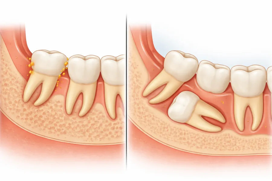
When a dentist informs you that your wisdom teeth are "impacted," it is easy to assume that all impactions are essentially identical. In clinical reality, impacted third molars exist along a wide anatomical spectrum. The clinical behavior, daily symptoms, and surgical requirements of a partially erupted wisdom tooth differ fundamentally from one that remains completely entombed beneath jawbone.
While partially impacted teeth tend to announce themselves through recurrent gum infections, foul odors, and localized swelling, fully impacted teeth often work in silence, quietly resorbing adjacent molar roots or developing expansive fluid-filled cysts. At Oral & Facial Surgery of Miami, Dr. Johanny Caceres uses advanced three-dimensional volumetric imaging to evaluate the exact depth, angulation, and health risks of every third molar, ensuring you receive timely, conservative, and definitive care.
1. The Impaction Spectrum: Defining Partial vs. Complete Bony Impaction
Third molars are the last teeth to calcify and erupt, usually attempting to emerge between the ages of 17 and 25. Because modern jaw arches frequently lack the retromolar space required for four full-sized molars, the erupting tooth encounters physical barriers that halt its forward trajectory. Maxillofacial surgeons classify these impactions based on the type of tissue obstructing the tooth and its depth within the mandible or maxilla.
Partially Impacted Wisdom Teeth: A tooth is classified as partially impacted when a portion of its enamel crown has pierced through the oral mucosa, while the remainder remains trapped beneath a dense flap of gum tissue (soft tissue impaction) or partially encased in bone (partial bony impaction). Because the protective seal of the oral mucosa is broken, the oral cavity has direct communication with the deeper follicle around the neck of the tooth.
Fully Impacted Wisdom Teeth: A fully impacted wisdom tooth has never broken through the gumline. It remains completely encapsulated within the jawbone (complete bony impaction) or submerged beneath thick, unbroken fibrous tissue. These teeth are frequently oriented at extreme angles, either horizontally parallel to the jawline, mesially tilted into the root of the second molar, or distally angled backward into the ascending ramus of the lower jaw.
| Clinical Feature | Partially Impacted Wisdom Teeth | Fully Impacted Wisdom Teeth |
|---|---|---|
| Visual Presentation | One or two cusps visible in the mouth; surrounded by a loose, inflamed flap of gum tissue (operculum). | Completely invisible to the naked eye; covered entirely by intact gingiva and cortical bone. |
| Primary Symptom Profile | Acute localized pain, swelling, bleeding on brushing, foul taste, and limited mouth opening (trismus). | Frequently asymptomatic for years; deep jaw pressure, radiating temporal tension, or zero outward pain. |
| Dominant Clinical Risk | Recurrent bacterial pericoronitis, severe dental decay on the neighboring molar, and periodontal bone loss. | Dentigerous cyst formation, internal root resorption of the second molar, and progressive jawbone thinning. |
| Surgical Technique | Conservative flap reflection; targeted bone relief; tooth elevation and primary wound closure. | Full surgical osteotomy, precise micro-sectioning of the tooth crown and roots, or elective coronectomy. |
2. Pathology and Symptoms: Bacterial Infection vs. Structural Destruction
The biological consequences of these two impaction states create distinct diagnostic challenges. One demands immediate attention due to acute pain, while the other requires vigilant clinical screening to prevent unseen structural damage.
The Problem with Partial Impaction (Pericoronitis): The loose mucosal flap draping over a partially erupted molar acts as a one-way valve. Microscopic food debris, plaque, and anaerobic oral bacteria easily slide beneath this operculum during chewing, but standard toothbrushes and floss cannot reach underneath to cleanse it. This triggers acute pericoronitis, a painful infection characterized by throbbing gum tissue, purulent discharge, bad breath, and severe jaw stiffness. If an opposing upper tooth bites down repeatedly on the swollen flap, the trauma accelerates the spread of infection toward the throat and neck.
The Silent Danger of Full Impaction (Cysts and Root Erosion): Because a fully impacted tooth has no exposure to oral saliva or food, it rarely develops surface bacterial infections. However, the microscopic developmental sac (follicle) that surrounds the crown of an unerupted tooth remains biologically active. Over years, this sac can accumulate fluid and expand into a dentigerous cyst. As the cyst expands, it quietly hollows out large portions of the mandibular jawbone, displacing adjacent teeth and weakening the jaw. Additionally, a fully impacted tooth wedged against the roots of a second molar can trigger osteoclasts that literally dissolve the healthy tooth roots from the outside in.
3. Precision Diagnostics: Why 3D CBCT Imaging Is Non-Negotiable
Traditional two-dimensional panoramic X-rays provide a general overview of dental arches, but they flatten complex 3D anatomy into a single distorted plane. When deciding whether an impacted tooth can safely remain in place or requires surgical extraction, 2D imaging often fails to show critical structural nuances.
At Oral & Facial Surgery of Miami, Dr. Johanny Caceres utilizes low-dose Cone Beam Computed Tomography (CBCT). This 3D imaging technology constructs a detailed volumetric map of your skull and jaws with sub-millimeter spatial resolution:
- Mapping the Inferior Alveolar Nerve: CBCT imaging reveals whether the roots of a deeply impacted lower molar sit beside, encircle, or press directly into the sensory nerve canal, virtually eliminating nerve injury risks during surgery.
- Second Molar Root Resorption: 3D scans detect early resorption craters on the backside of adjacent molars long before damage penetrates the dental pulp or causes clinical symptoms.
- Maxillary Sinus Proximity: For upper wisdom teeth, 3D imaging maps the delicate bony floor of the maxillary sinus, guiding the surgeon away from sinus perforation during extraction.
4. Surgical Solutions and What to Expect in Coral Gables
Whether your wisdom teeth are partially erupted or buried deep within the bone, modern oral and maxillofacial surgery provides a completely pain-free, comfortable treatment experience. At our Coral Gables surgical center, procedures are tailored precisely to the patient's anatomical complexity and comfort preferences.
Under soothing intravenous (IV) sedation, you drift into a peaceful twilight sleep, retaining zero memory of the surgery and feeling no discomfort whatsoever. For partially impacted teeth, Dr. Caceres gently reflects the soft tissue and elevates the tooth with minimal disturbance to surrounding structures. For complex, deeply impacted teeth, Dr. Caceres utilizes microsurgical sectioning, dividing the tooth crown and roots into small segments so they can be removed smoothly without excessive bone removal.
If a fully impacted tooth is intimately fused with the inferior alveolar nerve, Dr. Caceres can perform a protective coronectomy, cleanly removing the impaction-causing crown while leaving the harmless roots safely undisturbed. If you are experiencing gum soreness, jaw stiffness, or want to know whether your wisdom teeth require attention, contact Oral & Facial Surgery of Miami at (305) 552-1193 or schedule your consultation online today.