
Common Facial Fractures Explained: Orbital, Nasal & More
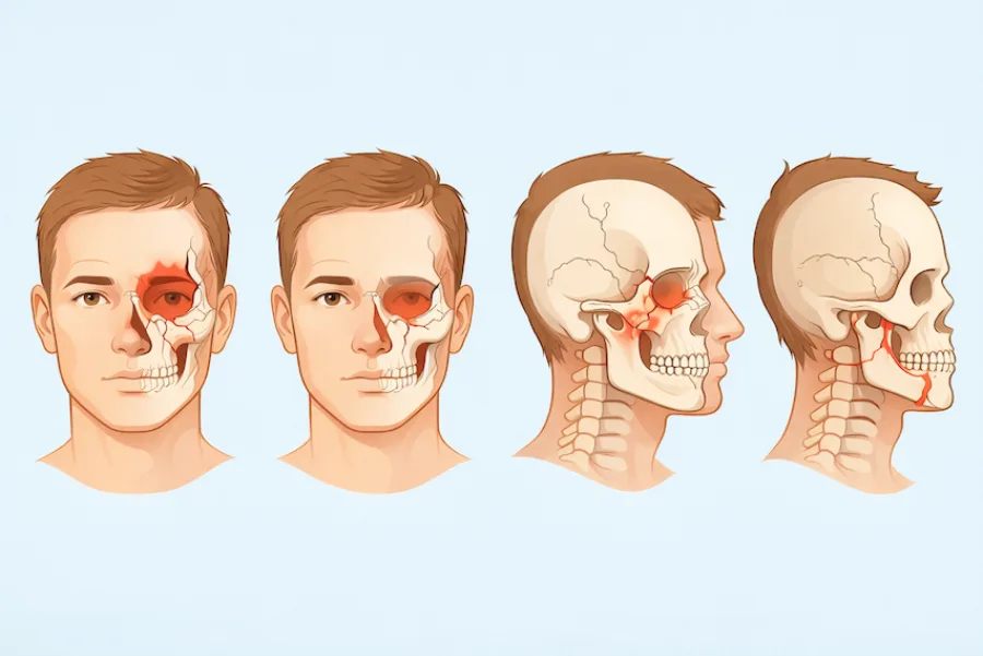
When people think of broken bones, they typically envision a casted forearm or a fractured ankle. Yet the human face contains an intricate architecture of fourteen delicate bones that support vision, breathing, mastication, and expressive communication. High-impact sports collisions, motor vehicle accidents, accidental falls, and physical altercations can shatter these fragile skeletal buttresses. Because the bones of the face surround vital structures like the eyes, airway, and brain, facial fractures are far more complex than standard orthopedic injuries.
Understanding the anatomical classifications of facial fractures, recognizing clinical red flags, and knowing how oral and maxillofacial surgeons reconstruct the facial skeleton ensures you receive timely, sight-saving and function-restoring care.
1. Anatomical Classifications: From Orbital Blowouts to ZMC Fractures
The facial skeleton functions as a biological crumple zone designed by nature to absorb traumatic energy and protect the neurocranium and brain. Fractures are classified based on the specific anatomical buttresses involved:
- Orbital Floor Blowout Fractures: The eye rests inside a bony cone. When an object larger than the orbital rim (such as a baseball, fist, or steering wheel) strikes the eye, hydraulic pressure pushes the eyeball backward, causing the paper-thin floor of the orbit (the maxillary roof) or medial wall (lamina papyracea) to buckle downward into the maxillary sinus. This can entrap the inferior rectus muscle, preventing the patient from looking upward and causing double vision (diplopia). In pediatric patients, a "white-eyed blowout fracture" can trap muscle without visible bruising, triggering severe nausea and dangerous slow heart rates (oculocardiac reflex) that require emergency surgical release.
- Zygomaticomaxillary Complex (ZMC) Fractures: Commonly known as tripod or tetrapod fractures, these involve the cheekbone separating at its suture lines with the forehead, upper jaw, skull base, and temporal bone. Hallmarks include a flattened cheek prominence, numbness in the upper lip from infraorbital nerve compression, and painful inability to open the mouth (trismus) when the depressed bone impinges on the lower jaw's coronoid process.
- Nasal and Naso-Orbito-Ethmoid (NOE) Fractures: Nasal bones are the most frequently broken bones in the face. When high-velocity impact drives the nasal bridge backward into the ethmoid sinus, it produces an NOE fracture. This can detach the medial canthal tendon that holds the inner corner of the eyelid, causing the eyes to appear unnaturally far apart (traumatic telecanthus).
- Mandibular Fractures: Because the lower jaw is shaped like a horseshoe, trauma to one side frequently causes a second fracture on the opposite side (such as a parasymphysis fracture combined with a contralateral condylar fracture). Patients present with sudden bite misalignment and severe pain when closing their teeth.
- Le Fort Midface Fractures: Described by French surgeon René Le Fort, these represent severe craniofacial trauma categorized into three levels: Le Fort I (horizontal detachment of the tooth-bearing upper jaw), Le Fort II (pyramidal midface fracture involving the nose and orbital rims), and Le Fort III (complete craniofacial disjunction, separating the facial skeleton from the skull base).
2. Clinical Red Flags, 3D Imaging, and Emergency Triage
Not every facial injury requires operating room reconstruction, but accurate diagnostic triage determines whether surgery is mandatory to preserve vision, airway, or dental occlusion.
| Fracture Type | Key Anatomical Structures | Signature Symptoms | Primary Treatment Approach |
|---|---|---|---|
| Orbital Blowout | Orbital floor, medial orbital wall, inferior rectus muscle. | Vertical double vision, sunken eye (enophthalmos), cheek numbness. | Transconjunctival exploration, muscle release, pre-formed titanium mesh reconstruction. |
| ZMC (Cheekbone) | Malar prominence, zygomatic arch, infraorbital rim. | Malar flattening, restricted jaw opening (trismus), subconjunctival hemorrhage. | Open reduction internal fixation (ORIF) with microplates at buttress points. |
| Nasal & NOE | Nasal bones, ethmoid sinus, medial canthal tendon. | Severe epistaxis, widened eye spacing (telecanthus), clear CSF rhinorrhea. | Closed or open reduction, transnasal canthopexy wiring, splinting. |
| Mandibular & Le Fort | Lower jaw corpus/condyles, maxillary alveolar ridge, midface buttresses. | Altered dental bite (malocclusion), intraoral bleeding, mobile facial skeleton. | Maxillomandibular fixation (MMF), rigid osteosynthesis with titanium mini-plates. |
Standard two-dimensional dental X-rays cannot fully visualize three-dimensional facial trauma. The gold standard diagnostic imaging is high-resolution, fine-slice (0.5 mm to 1.0 mm) maxillofacial Computed Tomography (CT) with 3D multiplanar reconstructions. These digital scans allow the facial trauma surgeon to examine the depth of displaced orbital fragments, measure orbital volume loss, and evaluate internal bone buttresses before entering the operating room.
3. Open Reduction Internal Fixation (ORIF) and Surgical Principles
The core philosophy of modern maxillofacial reconstruction is Open Reduction Internal Fixation (ORIF). This technique restores the three-dimensional skeletal pillars (vertical and horizontal buttresses) that establish facial height, facial width, and anterior projection.
Invisible Surgical Approaches: Oral and maxillofacial surgeons take pride in hiding surgical access points so patients have minimal or no visible external facial scars. Orbital floors are accessed through a transconjunctival incision placed on the inside of the lower eyelid. Upper and lower jaw fractures are exposed entirely through intraoral incisions made along the gums (gingivobuccal sulcus). Even cheekbone reductions can be performed through intraoral and hidden eyebrow or hairline margins.
Titanium Osteosynthesis and Mesh: Once displaced fragments are mobilized back into anatomical alignment, they are secured with ultra-low-profile titanium mini-plates (1.0 mm to 2.0 mm) and self-drilling monocortical screws. In orbital floor blowouts, custom-contoured or pre-formed titanium mesh, porous polyethylene sheets, or resorbable membranes span the defect, restoring true orbital volume and elevating the eyeball to eliminate double vision.
4. Recovery, Complication Prevention, and Specialized Care with Dr. Johanny Caceres
Following facial fracture repair, primary bone healing requires 6 to 8 weeks. During this period, adherence to surgical instructions determines the permanence of your aesthetic and functional recovery:
- Strict Dietary Modifications: For jaw and midface fractures, patients follow a non-chew soft food protocol for 4 to 6 weeks to avoid exerting destructive masticatory forces across healing titanium plates.
- Sinus Precautions: If your injury involves the maxilla, cheekbone, or orbit, you must not blow your nose for four weeks. Intranasal pressure can force air through fracture lines into your eye socket or facial tissues, causing sudden vision compromise or infection.
- Protection Against Re-Injury: Athletes must wear custom-fabricated protective polycarbonate facial masks when returning to active training to shield delicate healing bone from secondary trauma.
Delaying treatment for facial trauma can result in permanent deformities: sunken eyes (enophthalmos), persistent double vision, chronic malocclusion, and facial asymmetry that requires complex secondary osteotomies to repair. If you or a family member has experienced acute facial trauma, evaluation by a specialist is critical.
At our practice, Dr. Johanny Caceres is a board-certified oral and maxillofacial surgeon extensively trained in complex facial trauma, maxillofacial reconstruction, and emergency surgical stabilization. Located in Coral Gables, our practice combines compassionate bedside care with advanced 3D diagnostics and surgical technology to reconstruct delicate facial structures and restore your natural appearance and bite function.
If you require evaluation for facial bone trauma, suspect a broken jaw, or need emergency guidance, read our guides on what counts as a dental emergency and facial swelling emergencies.
To schedule an evaluation or discuss facial reconstructive treatment, contact our Coral Gables surgical office immediately at (305) 552-1193 or request an appointment online through our consultation booking portal.