
Why Regular Dental X-Rays Are Essential for Wisdom Teeth
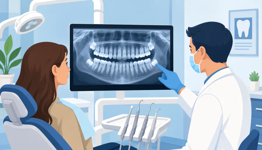
Third molars develop deep within the jawbone beneath dense mucosal tissue. Because they are the last teeth to form, visual clinical examinations alone cannot reveal how they are growing, where their roots are directed, or whether they threaten neighboring structures. Regular diagnostic radiographic imaging provides the only reliable window into third molar development.
At Oral and Facial Surgery of Miami, board-certified oral and maxillofacial surgeon Dr. Johanny Caceres relies on state-of-the-art diagnostic imaging to evaluate wisdom teeth. Routine radiographic tracking identifies silent complications early, protecting your healthy teeth and ensuring proactive, predictable surgical care.
The Limits of Visual Checks: Why Surface Exams Miss Impactions
During a routine dental checkup, a dentist looks at the surface of your teeth and gums with a mirror and probe. However, completely impacted wisdom teeth remain buried entirely below the gumline and inside the alveolar bone. Even partially erupted teeth only present a fraction of their enamel crown.
Without X-rays, serious biological complications develop undetected. Roots can curve directly into the mandibular nerve canal, un-erupted dental follicles can expand into destructive cysts, and horizontal crowns can erode adjacent second molar roots without triggering any early pain. Waiting for symptoms before taking an X-ray often means discovering damage after it has become irreversible.
| Imaging Modality | Dimensional View | Diagnostic Capability | Clinical Application |
|---|---|---|---|
| Bitewing Radiographs | 2D Flat Small View | Shows crowns of erupted teeth only | Detects interproximal cavities; cannot see impacted third molars |
| Panoramic X-Rays | 2D Wide Arch View | Shows all 32 teeth, sinus floor, and jaw borders | Excellent preliminary screening for impaction presence |
| 3D Cone Beam CT (CBCT) | 3D Volumetric Slices | Sub-millimeter cross-sectional depth of bone and nerves | Gold standard for high-risk surgical mapping and coronectomy planning |
2D Panoramic Radiographs vs 3D Cone Beam CT Volumetric Scans
For decades, the standard panoramic radiograph (panorex) was the primary tool for wisdom tooth assessment. While panoramic films provide a broad overview of the upper and lower dental arches, they remain two-dimensional representations of three-dimensional anatomical structures.
Because 2D images flatten structures, a wisdom tooth root and the inferior alveolar nerve canal often appear to overlap, even when they sit millimeters apart. To eliminate this ambiguity, Dr. Caceres utilizes 3D Cone Beam Computed Tomography (CBCT). A 3D scan allows the surgeon to rotate your jaw digitally, evaluating the precise thickness of cortical bone, root curvature, and exact spatial relationship to the nerve canal, ensuring maximum surgical safety.
Detecting Silent Pathology: Cysts, Caries, and Bone Erosion
Regular imaging is essential because severe wisdom tooth complications are predominantly asymptomatic during their early phases. As an angled third molar pushes against the adjacent second molar, it creates an un-cleanable biological niche. Radiographs detect cervical caries rotting the neighboring tooth root before nerve pain occurs.
In addition, un-erupted third molars are encased in a dental follicle that can expand into a dentigerous cyst. Radiographs reveal radiolucent shadows indicating fluid accumulation and internal bone resorption. Identifying cysts early allows for conservative enucleation before mandibular bone weakens to the point of fracture.
Advanced Digital Diagnostic Protocols at OFS Miami
At our practice, our surgical suite is equipped with modern, low-dose digital imaging systems. We prioritize diagnostic accuracy while minimizing radiation exposure to our patients.
During your consultation, Dr. Johanny Caceres reviews your 3D digital scans with you on high-definition displays, explaining anatomical landmarks and personalized treatment options. Whether monitoring developing teeth or planning gentle removal under outpatient IV twilight sedation, our advanced diagnostics ensure your care is safe, precise, and completely transparent.
Schedule your comprehensive 3D wisdom tooth evaluation today. Contact Oral and Facial Surgery of Miami at (305) 552-1193 or book your appointment online today.