
How Wisdom Teeth Can Cause Sinus Problems
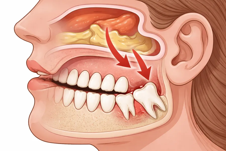
When chronic sinus congestion, facial pressure, or throbbing pain beneath the cheekbones persists despite nasal sprays and decongestants, patients rarely suspect their teeth. However, the upper dental arch and the respiratory system share an intimate anatomical border. The roots of your maxillary third molars (upper wisdom teeth) develop directly beneath, and frequently protrude into, the floor of the maxillary sinuses. When these teeth become impacted, infected, or horizontally angled, they can trigger profound sinus dysfunction.
At Oral & Facial Surgery of Miami, board-certified oral and maxillofacial surgeon Dr. Johanny Caceres frequently treats patients who have spent months visiting primary care physicians or allergists for unresponsive sinus infections, only to discover that an impacted third molar is the true inciting culprit. Understanding how upper wisdom teeth affect the maxillary sinus cavity is essential for finding lasting relief.
1. Anatomical Proximity: Maxillary Sinuses and Upper Third Molars
The maxillary sinuses are air-filled chambers situated directly behind your cheekbones and above the roots of your upper premolars and molars. In human anatomy, the bony barrier separating the tips of upper wisdom tooth roots from the sinus cavity (the sinus floor) is exceptionally thin, often measuring less than one to two millimeters in thickness. In many individuals, the bone is naturally dehiscent, meaning the root tips are separated from the sinus cavity solely by the delicate Schneiderian membrane (the mucosal lining of the sinus).
As wisdom teeth develop between the late teenage years and mid-twenties, their elongating roots expand superiorly. If the upper jaw lacks adequate room for normal vertical eruption, the tooth pushes upward and outward against the posterior sinus wall. This direct mechanical expansion elevates the mucosal floor, narrowing the sinus chamber, irritating sensory receptors, and giving rise to persistent facial fullness and heaviness that worsens when bending forward.
2. Odontogenic Sinusitis vs. Rhinogenic Sinusitis: Spotting the Differences
Sinusitis is broadly classified into two categories based on origin: rhinogenic (originating from viral upper respiratory infections, allergic rhinitis, or nasal anatomical blockages) and odontogenic (originating from dental pathology). Clinical studies indicate that up to 40 percent of chronic unilateral maxillary sinus infections stem from underlying dental disease. Because symptoms overlap substantially, patients frequently misattribute dental sinus pain to seasonal Florida pollen, humidity, or common colds.
However, odontogenic sinusitis carries specific diagnostic hallmarks. While common colds cause bilateral congestion and generalized fatigue, dental-induced sinus complications are overwhelmingly unilateral, concentrating in the single sinus cavity directly above the affected molar. Furthermore, because oral bacteria differ substantially from standard respiratory pathogens, odontogenic infections frequently generate foul-smelling nasal drainage, an unpleasant taste in the mouth, and localized tenderness when pressing on the upper alveolar ridge.
| Clinical Feature | Rhinogenic Sinusitis (Allergy / Viral) | Odontogenic Sinusitis (Wisdom Tooth) |
|---|---|---|
| Symmetry of Symptoms | Typically bilateral (both nasal passages and cheeks) | Strictly unilateral (concentrated on the side of the impacted tooth) |
| Nature of Drainage | Clear and watery (allergies) or mucoid yellowish (viral) | Thick, discolored, and distinctly foul-smelling or bad-tasting |
| Dental Symptoms | Generalized mild upper teeth ache during heavy congestion | Localized posterior gum tenderness, pericoronitis, chewing pain |
| Response to Nasal Sprays | Temporary to moderate relief with antihistamines / fluticasone | Negligible or zero response; symptoms recur quickly |
| Definitive Cure | Decongestants, immunotherapy, or sinus irrigation | Surgical extraction and eradication of the dental infection source |
3. The Mechanics of Complications: Pericoronitis, Cysts, and Oroantral Communication
When an upper third molar only partially breaks through the gingival margin, a mucosal flap (operculum) covers a portion of the crown. This creates an inaccessible crevice where food debris and virulent oral anaerobes accumulate, precipitating acute pericoronitis. Because the root tips border the sinus floor, this active bacterial reservoir can travel through micro-perforations in the thin cortical plate, seeding a virulent bacterial colony directly into the sterile sinus cavity.
Additionally, impacted teeth retained inside the maxilla can develop dentigerous cysts (fluid-filled epithelial sacs surrounding the unerupted crown). As the cyst slowly expands over years, it resorbs the surrounding maxillary bone and compresses the sinus floor from beneath. In severe scenarios, untreated cysts can expand to occupy the majority of the maxillary sinus, causing facial asymmetry and chronic obstruction of the osteomeatal complex (the natural drainage pathway of the sinus).
Another critical surgical consideration is the risk of oroantral communication, an unnatural opening connecting the oral cavity to the maxillary sinus. When upper wisdom teeth with roots deeply anchored in the sinus floor are removed, an opening can occur if not managed with meticulous surgical finesse. As an oral and maxillofacial surgeon, Dr. Caceres applies specialized flap design, gentle tooth sectioning, and primary closure techniques to safeguard the sinus floor and prevent chronic fistula formation.
4. Precision 3D Diagnostics and Specialist Care at OFS Miami
Standard two-dimensional panoramic X-rays provide an overview of dental alignment, but they flatten complex 3D anatomical structures into a single plane. This makes it impossible to definitively determine whether a wisdom tooth root is merely overlapping the sinus or penetrating inside it. At Oral & Facial Surgery of Miami, Dr. Johanny Caceres utilizes in-office Cone Beam Computed Tomography (CBCT) to capture cross-sectional, sub-millimeter 3D images of your maxilla, roots, and sinus cavities.
This advanced diagnostic capability enables our team to evaluate mucosal thickening, pinpoint bone resorption, and map an ultra-precise surgical trajectory before beginning treatment. Removing problematic upper wisdom teeth relieves the mechanical strain against the sinus floor, halts the upward seeding of oral pathogens, and allows the Schneiderian membrane to regenerate naturally. For anxious patients, we offer customized IV sedation in our state-of-the-art Coral Gables facility, ensuring you remain completely comfortable and relaxed throughout your procedure.
If you are suffering from unrelenting sinus pressure, unilateral cheek discomfort, or recurring nasal infections that refuse to heal, your upper wisdom teeth may be the hidden cause. Contact Oral & Facial Surgery of Miami today at (305) 552-1193 or schedule your consultation online to receive the comprehensive evaluation and relief you deserve.