
3D Imaging: Revolutionizing Wisdom Tooth Treatment Planning
Third molar extraction is one of the most common surgical procedures performed in dentistry, yet no two dental arches present the same anatomical configuration. While traditional two-dimensional dental X-rays provided an adequate general overview for decades, modern maxillofacial surgery demands sub-millimeter precision. The integration of high-resolution three-dimensional imaging has established a new clinical benchmark for surgical planning, transforming complex impactions into highly predictable, minimally invasive outpatient procedures.
At Oral & Facial Surgery of Miami, board-certified oral and maxillofacial surgeon Dr. Johanny Caceres utilizes in-office Cone Beam Computed Tomography (CBCT) to evaluate third molars in three distinct spatial planes. Understanding how 3D diagnostics eliminate clinical blind spots provides anxious patients with profound reassurance before sitting in the surgical chair.
1. Beyond 2D Panoramics: The Science of Cone Beam Computed Tomography (CBCT)
Standard panoramic radiographs (panorex) flatten the curved, three-dimensional structure of the human maxilla and mandible into a single flat image. In this compressed format, anatomical structures naturally overlap. A panoramic film can show that a wisdom tooth root appears to cross over the mandibular nerve canal, but it cannot show whether the root is situated on the facial side of the nerve, on the tongue side, or if the nerve actually runs through a perforation within the root itself.
Cone Beam Computed Tomography overcomes these limitations by projecting a divergent cone-shaped beam of low-dose radiation through the patient while rotating 360 degrees around the head. Sophisticated reconstruction algorithms generate hundreds of high-resolution, volumetric cross-sections (axial, coronal, and sagittal views). Oral surgeons can slice through digital bone tissue at fractions of a millimeter, examining root dilacerations, cortical plate density, and surrounding soft tissue borders with absolute spatial clarity.
2. Mapping Critical Anatomy: Inferior Alveolar Nerves and Maxillary Sinus Floors
The primary hazard during lower wisdom tooth removal is neurovascular injury, specifically trauma to the inferior alveolar nerve (IAN), which provides sensation to the lower lip, chin, and front teeth. When 2D imaging reveals dark banding, deviation of the mandibular canal, or loss of the cortical canal line, the risk of close proximity is heightened. A 3D CBCT scan confirms the exact spatial relationship. If the nerve is compressed between divergent roots, Dr. Caceres can plan a coronectomy (intentional partial odontectomy) to remove the infected crown while leaving healthy root tips securely untouched, completely eliminating neurosensory deficit risks.
In the upper jaw, 3D imaging evaluates the maxillary sinus floor. Upper third molars frequently share a thin bony partition with the air-filled maxillary sinus cavity. By evaluating bone thickness and sinus membrane (Schneiderian membrane) mucosal health in 3D cross-sections, an oral surgeon can identify hidden cysts, plan gentle sectioning angles, and prevent post-surgical oroantral communications (unnatural openings between the mouth and sinus).
| Diagnostic Feature | Traditional 2D Panoramic Film | In-Office 3D CBCT Volumetric Scan |
|---|---|---|
| Spatial Dimensionality | Flat height and width; zero depth or volumetric thickness | Complete 3D spatial rendering (axial, coronal, sagittal cross-sections) |
| Nerve Canal Relationship | Shows visual overlap but cannot determine true spatial contact | Pinpoints whether nerve is buccal, lingual, or grooved between roots |
| Sinus Floor Accuracy | Distorts sinus floor thickness due to projection angulation | Measures bone thickness and mucosal thickening to the sub-millimeter |
| Adjacent Root Health | Often obscures external root resorption on neighboring 2nd molars | Reveals early hidden root resorption craters on adjacent molars |
| Surgical Invasiveness | May require wider exploratory bone removal during surgery | Enables conservative, targeted osteotomy with minimal tissue trauma |
3. Preoperative Odontotomy and Custom Surgical Trajectories
Beyond risk reduction, 3D imaging changes how surgical extractions are executed. Rather than applying heavy elevation forces that stress the mandible, an oral surgeon uses 3D reconstructions to map an exact trajectory for tooth sectioning (odontotomy). Visualizing the internal curvature of each root allows the surgeon to plan precision cuts, separating the tooth crown from its roots or dividing multi-rooted molars before elevation.
This targeted engineering approach minimizes the amount of surrounding bone that must be removed (osteotomy). Because less bone is trimmed and minimal mechanical traction is transferred to the jawbone, patient recovery is substantially accelerated. Operating time is reduced, localized postoperative edema (swelling) is noticeably dampened, and trismus (jaw muscle stiffness) is minimized during the critical first 72 hours of healing.
4. Clinical Precision, Reduced Morbidity, and Care at Oral & Facial Surgery of Miami
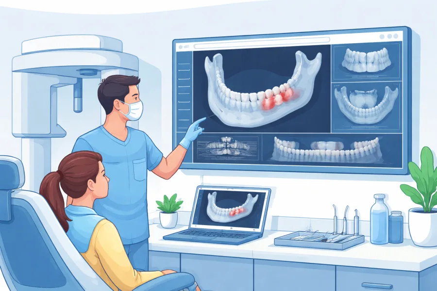
At Oral & Facial Surgery of Miami (OFS Miami), patient safety and comfort drive every clinical decision. During your initial consultation at our surgical center, Dr. Johanny Caceres reviews your 3D CBCT scans alongside you on high-definition chairside monitors. Walking you through the 3D volume helps clarify your anatomy, allowing you to see exactly where your wisdom teeth rest and why extraction is indicated.
Combined with customized outpatient IV sedation, our digital surgical planning ensures your procedure is comfortable, gentle, and swift. Knowing your surgeon has visualized every nerve, bone wall, and root curve beforehand turns a potentially stressful surgery into a smooth, reassuring healthcare experience.
If you or your teenager require wisdom tooth evaluation, experience the safety and confidence of state-of-the-art 3D diagnostics. Contact Oral & Facial Surgery of Miami today at (305) 552-1193 or schedule your consultation online to take the first step toward optimal oral health.